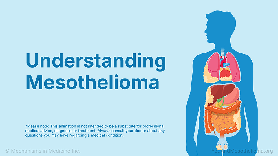
Understanding Mesothelioma — Animation
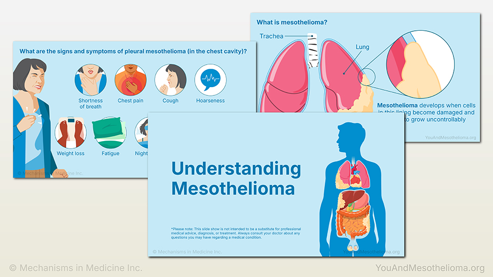
Understanding Mesothelioma — Slide Show
1. What is mesothelioma? What are the types?
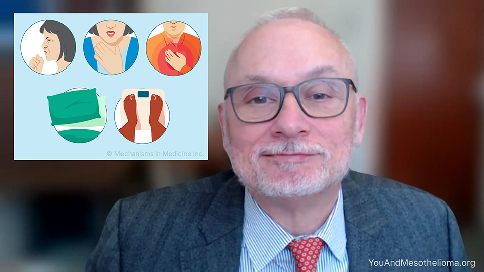
2. What are the signs and symptoms of mesothelioma?

3. How is mesothelioma diagnosed? What tests are used?
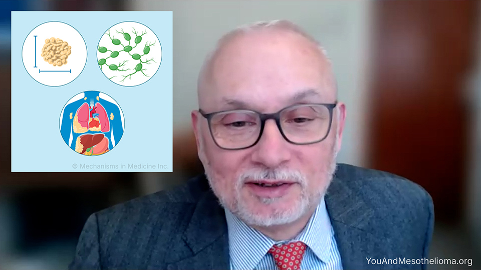
4. What are the stages of mesothelioma?
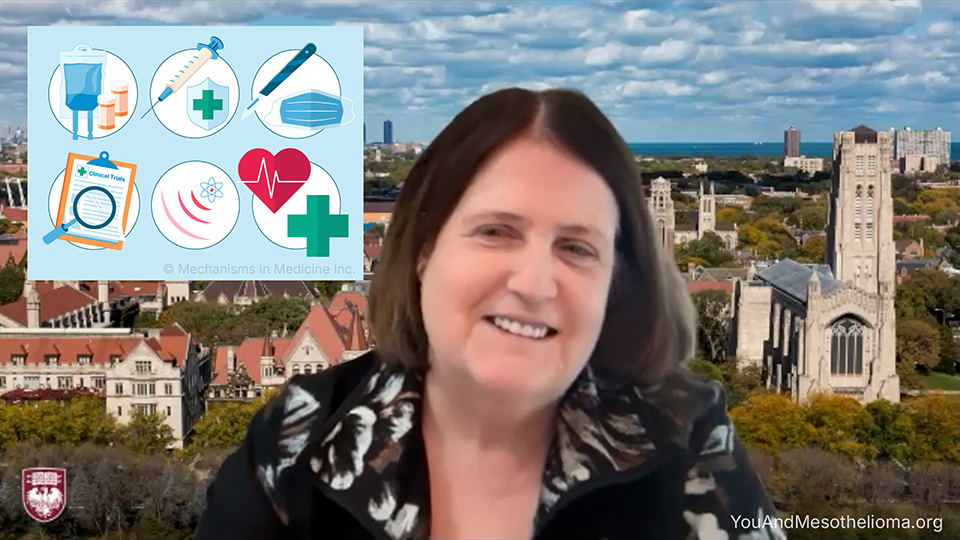
5. How is mesothelioma treated and managed?

6. Why is a mesothelioma specialist and multidisciplinary care team important for treatment?

7. Rosa's story: How did you find out you had mesothelioma?

8. Rosa's story: What challenges and successes have you had with your mesothelioma treatment?
9. Ben's story: How did you find out you had mesothelioma?

10. Ben's story: What challenges and successes have you had with your mesothelioma treatment?
How is mesothelioma diagnosed? What tests are used? — Expert Video
Dr. Hedy Lee Kindler, a medical oncologist and professor of medicine at the University of Chicago, explains how mesothelioma is diagnosed and what tests are used. For pleural mesothelioma, diagnosis begins with a physical exam and imaging tests such as a chest X-ray, CT scan, PET scan, and sometimes an MRI. If fluid is present around the lungs, it may be drained and tested using a procedure called a thoracentesis. However, a biopsy is usually needed to confirm the diagnosis, often done using image-guided techniques or a thoracoscopy, which allows doctors to take multiple tissue samples. For peritoneal mesothelioma, diagnosis also starts with a physical exam and imaging such as CT or MRI scans, and sometimes a PET scan. Fluid in the abdomen may be removed and analyzed using a procedure called a paracentesis. A biopsy is still required, either through image-guided needle sampling or a laparoscopy to collect tissue from the abdomen. In both pleural and peritoneal mesothelioma, germline and somatic testing may be done to look for molecular changes that help guide treatment decisions.
Click here to take our SURVEY
Your feedback is important to us! We will use your feedback to develop future areas of content about mesothelioma which will help other patients, caregivers, and families.
Understanding Mesothelioma — Animation
Understanding Mesothelioma — Slide Show
1. What is mesothelioma? What are the types?
2. What are the signs and symptoms of mesothelioma?
3. How is mesothelioma diagnosed? What tests are used?
4. What are the stages of mesothelioma?
5. How is mesothelioma treated and managed?
6. Why is a mesothelioma specialist and multidisciplinary care team important for treatment?
7. Rosa's story: How did you find out you had mesothelioma?
8. Rosa's story: What challenges and successes have you had with your mesothelioma treatment?
9. Ben's story: How did you find out you had mesothelioma?
10. Ben's story: What challenges and successes have you had with your mesothelioma treatment?